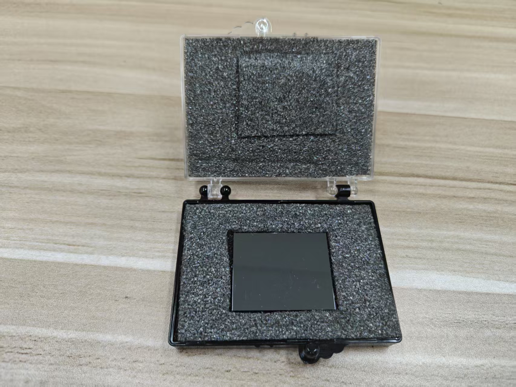
CZT-based detectors have emerged as a powerful and efficient technology for
medical imaging, particularly in applications like
Single Photon Emission Computed Tomography (SPECT) and
gamma imaging. These detectors offer significant advantages over traditional imaging technologies such as
scintillation-based detectors or
crystal-based detectors in terms of
energy resolution,
efficiency, and the ability to operate at
room temperature. Below is a detailed explanation of how
CZT-based detectors function in medical imaging, with a focus on the underlying principles, advantages, and specific applications in the field.
## 1. Basic Working Principle of CZT Detectors
The working principle of
CZT-based detectors in medical imaging is based on the interaction between
gamma radiation (or
X-rays) and the CZT semiconductor material. The basic steps involved in the detection process are:
## a. Photon Interaction
* When
gamma photons (or
X-rays) from the patient’s body interact with the
CZT detector, the energy from the photon is absorbed by the material, which leads to the generation of
electron-hole pairs within the semiconductor. This interaction typically happens through the
photoelectric effect or
Compton scattering.
## b. Charge Carrier Generation
* The energy from the incident photon excites electrons in the CZT crystal, causing them to jump from the
valence band to the
conduction band, leaving behind a
hole in the valence band. The
electron-hole pairs created in this process represent the energy deposited by the photon in the material.
## c. Charge Collection
*
Electrodes are applied to the
CZT crystal to create an electric field that helps separate the
electrons and
holes. The
electrons are attracted to the
anode, and the
holes are attracted to the
cathode. This movement of charge carriers under the influence of the electric field leads to the formation of a
charge pulse.
## d. Signal Detection
* The
charge pulse is collected by the
electrodes and passed through a
charge-sensitive preamplifier that converts the charge into a voltage signal. This signal is then amplified and processed by
electronics in the imaging system.
## e. Energy and Position Determination
* The
energy of the incident photon is determined by measuring the magnitude of the charge pulse, which is directly proportional to the energy deposited in the CZT crystal. The
position of the photon interaction within the crystal is typically determined using
position-sensitive detectors, such as
position-sensitive photodiodes or
array-based detectors that can localize the
interaction point.
This process allows CZT-based detectors to convert
gamma radiation into an
electrical signal, which is subsequently processed to create
detailed images for medical diagnostics.
## 2. Advantages of CZT-Based Detectors in Medical Imaging
CZT detectors provide several key advantages that make them particularly well-suited for
medical imaging applications such as
SPECT and
gamma cameras:
## a. High Energy Resolution
* One of the most significant benefits of
CZT-based detectors in medical imaging is their
excellent energy resolution. Energy resolution refers to the detector's ability to distinguish between
different photon energies. CZT typically offers an energy resolution of
5-10% FWHM at
662 keV (the energy of the Cesium-137 gamma line), which is far superior to many other detector technologies like
scintillators or
silicon-based detectors.
*
High energy resolution is essential in medical imaging, especially in
SPECT, where distinguishing between different energies from the radiopharmaceuticals used for imaging is critical for accurate
quantification and
differentiation of tissues, tumors, or other anomalies.
## b. Room Temperature Operation
* Unlike
germanium detectors, which require cryogenic cooling,
CZT detectors can operate effectively at
room temperature. This eliminates the need for complex and expensive
cooling systems, which makes the equipment more
cost-effective and
easier to maintain. For medical imaging, where compact and user-friendly equipment is essential, the
room temperature operation of CZT simplifies the design and improves the practicality of portable
gamma cameras and
SPECT systems.
## c. High Detection Efficiency
* The high
atomic number (Z = 48 for cadmium and Z = 52 for tellurium) of
CZT leads to increased
photon interaction cross-sections for
gamma rays and
X-rays, resulting in
high detection efficiency. This is particularly beneficial in
SPECT, where high photon fluxes are often involved, as CZT detectors can achieve better
detection efficiency at smaller
thicknesses compared to scintillation detectors.
* The
high density of CZT (5.85 g/cm³) ensures that more of the
incident radiation is absorbed, making the detector more sensitive to lower activity levels of radiopharmaceuticals used in medical diagnostics.
## d. Compact and Lightweight
* CZT-based detectors can be fabricated into
compact,
lightweight devices, making them ideal for
portable and
mobile medical imaging systems. This is especially useful in
point-of-care applications where rapid, bedside diagnostics are required, or in environments where traditional
hospital-based imaging systems are not available or practical.
* The
small form factor allows for easier integration into handheld or portable gamma cameras, providing
greater flexibility in use. For instance,
SPECT systems can be made more
portable, enabling quicker imaging and diagnosis in
emergency or
field-based medical settings.
## e. Resistance to Radiation Damage
*
CZT detectors exhibit good
radiation hardness, which means they are more resistant to
radiation-induced damage compared to other materials like
silicon. This is important in
medical imaging, where the detectors are exposed to
high levels of radiation over time. The long-term
stability and
durability of CZT make it a reliable choice for continuous
medical imaging in clinical settings.
## 3. Applications of CZT in Medical Imaging
CZT-based detectors are particularly useful in several key
medical imaging modalities, including:
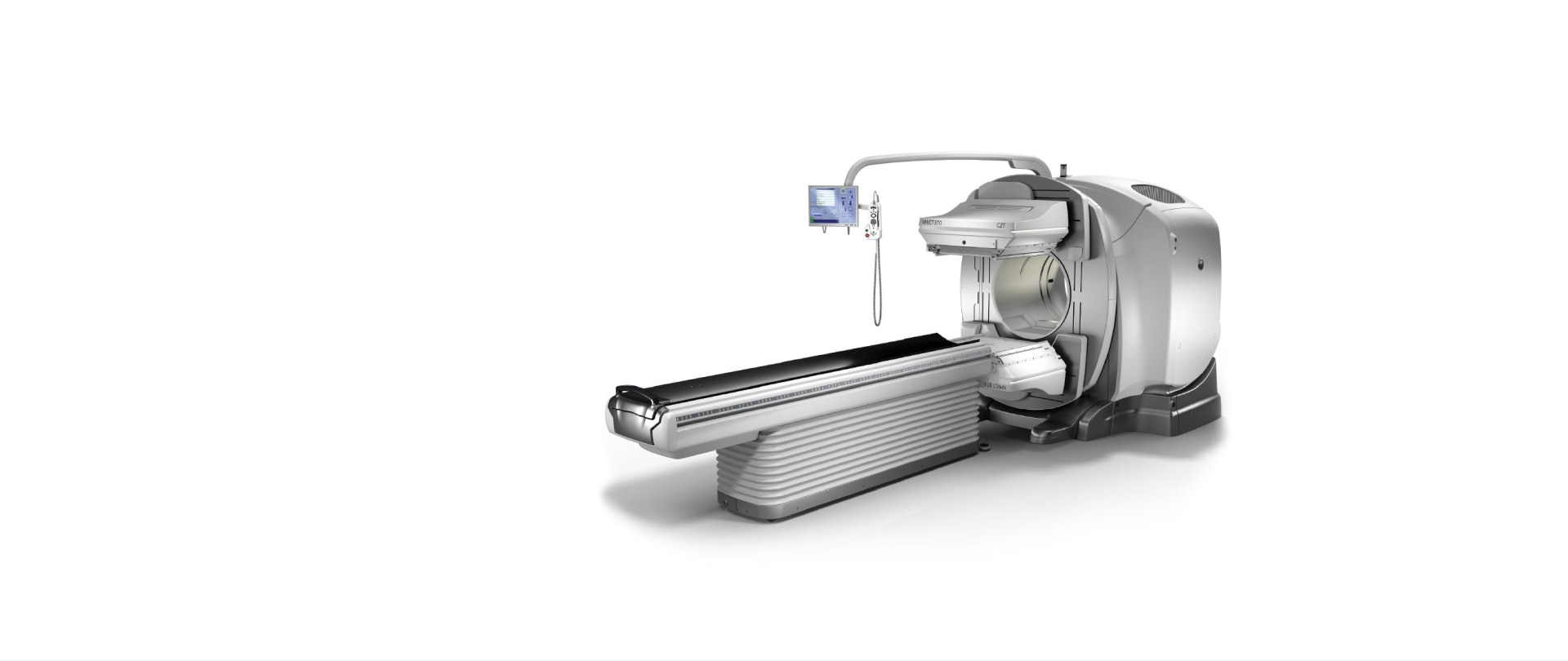
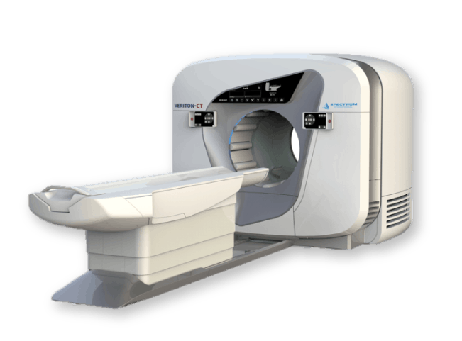
## a. Single Photon Emission Computed Tomography (SPECT)
*
SPECT is a type of
nuclear imaging that provides detailed,
3D images of the distribution of a
radiopharmaceutical within the body. The radiopharmaceutical emits
gamma rays, which are detected by the CZT-based detectors. The
high energy resolution of CZT is crucial in
SPECT for accurately differentiating the energies of
gamma photons, improving the image quality and providing more precise
diagnostic information.
* CZT-based
SPECT systems can achieve better
image resolution and
contrast than traditional systems, improving the detection and quantification of
tumors,
heart disease, and other medical conditions.
## b. Gamma Cameras
*
Gamma cameras are commonly used in both
SPECT and
general nuclear medicine imaging. They detect
gamma radiation emitted from the body after the administration of a radiopharmaceutical. CZT detectors are used in these cameras to offer
higher resolution and
faster imaging compared to traditional
scintillation-based detectors. The
compactness of CZT-based cameras makes them more adaptable to
small spaces and
emergency scenarios.
## c. Positron Emission Tomography (PET) / PET/CT Imaging
* Although
CZT detectors are typically associated with
SPECT, they are also explored for use in
positron emission tomography (PET). In this case, CZT detectors can be integrated into
PET scanners to improve the
time resolution and
detection efficiency, especially when paired with
CT imaging for
combined PET/CT scans.
## d. Portable and Point-of-Care Imaging
* The ability of CZT detectors to function at room temperature and their high
energy resolution make them highly suitable for
portable medical imaging devices. In
field settings,
emergency medicine, or
immediate diagnostic settings, handheld CZT detectors can be used for
quick screening of radiation in the body, particularly for
cancer or
cardiac assessments.
## 4. Challenges and Considerations
While CZT-based detectors offer many advantages, there are some challenges that need to be addressed in the context of medical imaging:
## a. Cost and Manufacturing Complexity
* The production of high-quality
CZT crystals can be complex and expensive, making the detectors costlier than alternative technologies such as
scintillation-based detectors. This cost can be a limiting factor for widespread adoption in some clinical settings, especially for
budget-conscious hospitals or
healthcare systems.
## b. Material Defects and Crystal Quality
* The performance of
CZT detectors is highly dependent on the
quality of the crystals.
Defects in the crystal lattice can result in
lower charge collection efficiency,
degraded energy resolution, and
reduced sensitivity. Ensuring high-quality crystals is critical for achieving the best imaging performance, but the process of growing defect-free CZT crystals remains a technical challenge.
## Conclusion
CZT-based detectors offer significant advantages for
medical imaging, particularly in
SPECT and
gamma imaging applications. Their
high energy resolution,
room temperature operation,
high detection efficiency, and
compact form factor make them highly effective for
portable,
high-resolution imaging. While there are challenges associated with
cost and
material quality, the superior performance of CZT in
nuclear medicine and its ability to operate in compact, mobile systems position it as an ideal choice for
cutting-edge medical imaging.
CdZnTe Association (CdZnTe.com)
https://www.cdznte.com/blog/how-do-czt-based-detectors-work-in-medical-imaging.html