## Introduction
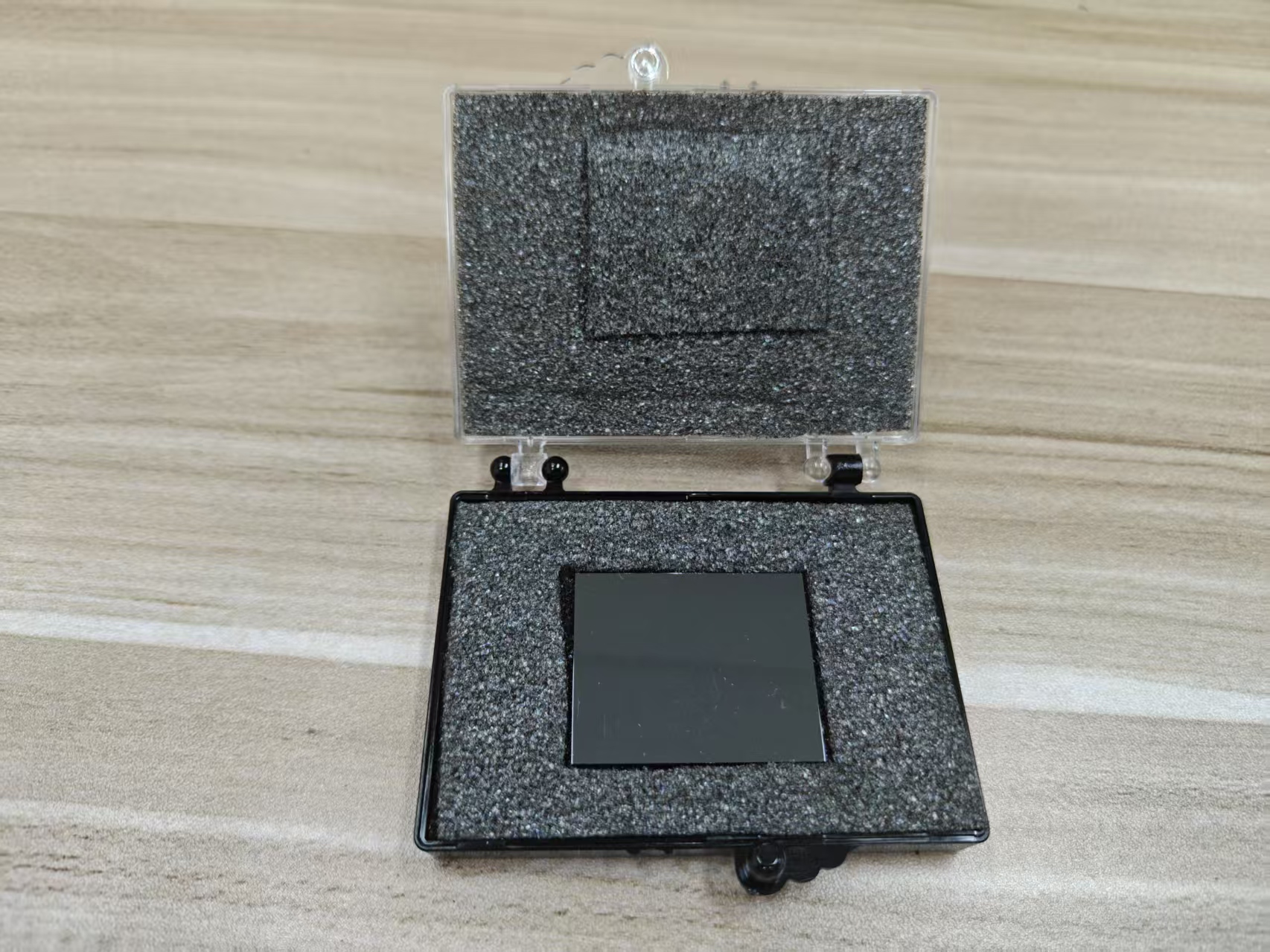
Characterizing the passivation layers in CdZnTe (
Cadmium Zinc Telluride) detectors is essential for understanding how surface chemistry and structure affect electrical performance, particularly leakage current, interfacial stability, and long-term reliability. Two of the most powerful techniques for such nanoscale investigations are
Transmission Electron Microscopy (TEM) and
Energy-Dispersive X-ray Spectroscopy (EDS). When used in combination, these tools provide a comprehensive picture of both the
structural morphology and
elemental composition of the passivation layers, including information on layer thickness, crystallinity, interdiffusion, and chemical uniformity. Their synergistic application enables precise evaluation of interfacial phenomena, thin-film uniformity, and passivation efficacy in Pt-CdZnTe and other detector configurations.
## Role of Transmission Electron Microscopy (TEM)
TEM provides high-resolution imaging of material cross-sections, capable of resolving features at atomic or near-atomic scale. In the context of CdZnTe passivation layers, TEM enables detailed visualization of:
## Layer Thickness Measurement
TEM is particularly effective in determining the physical thickness of passivation films. In cross-sectional mode:
* The
contrast between different layers (e.g., CdZnTe substrate, native oxide, intentional passivation, and electrode) allows for precise thickness delineation.
* High-resolution TEM (HRTEM) can resolve
ultra-thin layers down to a few nanometers, making it ideal for analyzing atomic-scale surface passivation.
* For multilayer structures (e.g., oxide + nitride), each individual layer's thickness can be separately measured.
## Interface Quality and Roughness
TEM reveals the
sharpness or gradation of interfaces:
* A
sharp, well-defined interface suggests minimal interdiffusion and a stable, effective passivation process.
* A
diffuse or rough interface indicates possible chemical reactions, mechanical stress, or poor deposition control, which can lead to increased trap density and degraded electronic behavior.
## Crystallinity and Amorphous Structure
TEM differentiates between crystalline and amorphous regions:
*
Amorphous passivation layers (e.g., SiO₂, Al₂O₃) show no lattice fringes in HRTEM and appear as uniform, structureless regions.
*
Crystalline secondary phases (e.g., CdTeO₃ or Te-rich inclusions formed during oxidation or annealing) can be detected by their distinct lattice patterns.
* Polycrystalline passivation layers, if present, are revealed by
grain boundaries and diffraction contrast, which can influence dielectric behavior.
## Defect Identification
TEM can reveal
microstructural defects, such as:
*
Dislocations or stacking faults at the interface
*
Void formation or delamination between passivation and substrate
*
Nanocracks introduced during thermal cycling or mechanical processing
Such defects are key indicators of poor thermal compatibility or deposition mismatch, which can compromise the long-term integrity of the passivation layer.
## Role of Energy-Dispersive X-ray Spectroscopy (EDS)
EDS is typically performed within a TEM or scanning TEM (STEM) system, allowing for simultaneous
elemental analysis and spatial resolution. In characterizing CdZnTe passivation layers, EDS is critical for:
## Elemental Composition Analysis
EDS provides
qualitative and quantitative data on the elemental constituents within the passivation region:
* Identifies elements such as
O, Si, Al, Zn, Cd, Te, Pt, and any trace contaminants (e.g., Cl, Na, or C).
* Distinguishes
native oxides (e.g., TeO₂ or CdO) from
intentional dielectric layers (e.g., Al₂O₃, Si₃N₄).
* Detects unwanted
diffusion of electrode metals (e.g., Pt migration into CdZnTe) that may occur during annealing or poor deposition control.
## Composition Uniformity and Depth Profiling
By conducting
line scans or elemental mapping across the interface:
* EDS can determine
composition gradients, which indicate interdiffusion between layers.
* A
sharp compositional transition reflects a stable and abrupt interface, whereas
gradual variation signals chemical reactivity or diffusion.
* Depth-resolved elemental profiles (e.g., from surface to bulk) reveal the
extent of passivation layer penetration, oxidation depth, or segregation of elements like Zn and Te.
## Detection of Secondary Phases
EDS is useful for identifying
localized chemical anomalies, such as:
*
Te-rich regions due to tellurium segregation near the surface
*
Cd-depleted zones near interface regions that might form during oxidation or etching
* Formation of
oxides or intermetallic compounds, particularly at the Pt/CdZnTe interface following thermal treatment
These secondary phases often correspond to performance-degrading features like trap sites or leakage paths, and their presence guides optimization of surface cleaning and passivation protocols.
## Correlated Analysis: TEM-EDS Integration
The true power of combining TEM and EDS lies in their
correlated analysis capability, providing both structural and chemical insights in a single experiment. This integration enables:
*
Thickness measurement with elemental verification, ensuring that observed contrast changes in TEM correspond to actual compositional boundaries.
*
Elemental mapping over structural defects, revealing how impurities or phase segregation aligns with dislocations, voids, or rough interfaces.
*
Time-resolved studies of annealed samples, showing how passivation evolves thermally in both structure and chemistry.
For example, a TEM-EDS study of a Pt-CdZnTe detector before and after annealing may reveal the growth of a 5–10 nm oxide layer, diffusion of Pt into the near-surface region, and depletion of Zn at the interface—all crucial indicators of interfacial reactivity and passivation failure.
## Application Examples
## Native Oxide Characterization
TEM can image the thin, spontaneous native oxide (~1–5 nm) that forms on CdZnTe after exposure to air. EDS confirms its composition, often detecting elevated Te and O levels, suggesting formation of TeO₂ or mixed oxides. These native oxides can introduce trap states and need to be removed or stabilized through chemical etching or controlled passivation.
## ALD or PECVD Passivation Films
For detectors passivated with Atomic Layer Deposition (ALD) or Plasma-Enhanced Chemical Vapor Deposition (PECVD), TEM verifies the
uniformity and conformality of the film across complex topography, while EDS validates that no unintended interfacial reactions or diffusion of passivation species (e.g., Al or Si) into the CdZnTe substrate has occurred.
## Interfacial Reactions with Metal Contacts
In Pt-contacted CdZnTe detectors subjected to high-temperature annealing, TEM-EDS can detect the formation of
intermetallic phases at the interface. For instance, localized EDS may reveal the presence of Pt-Te compounds at the boundary, indicating degradation of the Schottky barrier. Such insights are crucial for determining safe annealing conditions that preserve contact integrity.
## Conclusion
Transmission Electron Microscopy (TEM) and Energy-Dispersive Spectroscopy (EDS) are indispensable tools for the nanoscale characterization of CdZnTe detector passivation layers. TEM provides precise structural information such as layer thickness, interface morphology, crystallinity, and defect states, while EDS delivers complementary elemental analysis, identifying composition, uniformity, diffusion, and secondary phases. Together, these techniques enable a deep understanding of the physical and chemical nature of passivation layers, guiding the optimization of fabrication processes and ensuring high-performance, low-leakage, and spectrally stable radiation detectors. The inclusion of such advanced characterization techniques elevates the development of CdZnTe-based technologies to a more predictive, materials-informed level.
CdZnTe Association (CdZnTe.com)
https://www.cdznte.com/blog/how-do-transmission-electron-microscopy-tem-and-energy-dispersive-spectroscopy-eds-help-characterize-the-thickness-and-composition-of-cdznte-passivation-layers.html