## Introduction to Te Inclusion in CZT Material
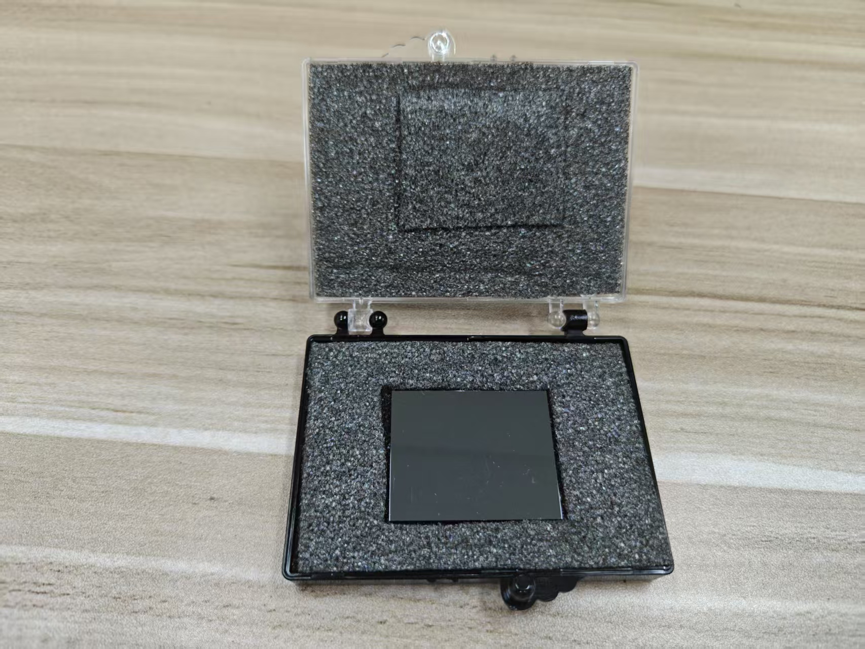
Cadmium Zinc Telluride (CZT) is a widely used semiconductor for photon-counting computed tomography (PCCT) due to its high atomic number, wide bandgap, and direct conversion of x-rays to charge carriers. However, CZT crystals often contain microscopic tellurium (Te) inclusions—metallic Te precipitates trapped within the crystal lattice during growth. These inclusions act as localized defects that perturb charge transport and impact detector performance. In PCCT imaging, where photon-counting precision and energy resolution are essential, the density, size, and distribution of Te inclusions play a critical role in defining charge collection efficiency, spectral fidelity, and ultimately image quality.
## Formation and Characterization of Te Inclusions
Te inclusions arise during the Bridgman or high-pressure Bridgman growth processes commonly used for CZT crystal production:
*
Supersaturation and Segregation: During solidification, Te tends to segregate due to differences in solubility between the solid and liquid phases, forming micron-scale inclusions.
*
Size and Distribution: Inclusion size ranges from sub-micron to several microns, and distribution may be random or clustered along growth directions.
*
Detection and Quantification: Optical microscopy, infrared imaging, and x-ray topography are used to map inclusion density, which can vary from a few per cubic centimeter to hundreds in high-volume crystals.
Understanding inclusion density is essential because it correlates directly with charge transport anomalies and spatial non-uniformities in detector response.
## Impact on Charge Transport
Te inclusions disrupt the uniform drift of electrons and holes generated by photon interactions in CZT:
*
Carrier Trapping and Recombination: Te inclusions introduce deep-level states that trap electrons or holes. Trapped carriers do not contribute to the measured pulse, reducing charge collection efficiency (CCE).
*
Local Electric Field Distortion: Metallic Te precipitates perturb the electric field in the detector, causing local variations in carrier drift velocity. This leads to pulse-height variations and broadened spectral peaks.
*
Increased Charge Sharing: Near-pixel field distortion can redirect a portion of charge to neighboring pixels, exacerbating charge-sharing effects and degrading spatial resolution.
*
Pulse Tail Formation: Partial trapping and delayed release of carriers from inclusion sites generate low-energy pulse tails, reducing energy resolution and causing spillover between energy bins in multi-threshold PCCT detectors.
These mechanisms collectively reduce the fidelity of energy measurement and compromise quantitative material decomposition.
## Effects on Energy Resolution and Spectral Imaging
Energy resolution is particularly sensitive to Te inclusion density:
*
Spectral Broadening: Trapped carriers and local field perturbations broaden photon energy peaks, decreasing the ability to distinguish adjacent energy bins.
*
Low-Energy Tails: Partial charge collection from interactions near inclusions creates extended low-energy tails, contaminating lower-energy bins and reducing the accuracy of material decomposition.
*
Threshold Misclassification: In multi-energy threshold systems, pulses affected by inclusions may fall below intended thresholds or be misassigned, directly impacting virtual monoenergetic image quality.
High inclusion density correlates with lower energy resolution and diminished spectral separation, reducing contrast and quantitative reliability in PCCT imaging.
## Influence on Spatial Resolution and Image Uniformity
Te inclusions also affect the spatial characteristics of PCCT images:
*
Localized Non-Uniform Response: Pixels overlying regions of high inclusion density exhibit lower or more variable counts due to reduced CCE, introducing streaks or mottling in reconstructed images.
*
Charge Diffusion Artifacts: Distorted drift paths can cause charge to spread laterally, blurring small structures in high-resolution imaging.
*
Small-Pixel Effects: While small pixels mitigate hole trapping effects, they are more sensitive to local electric field distortions from inclusions, making pixel-to-pixel uniformity highly dependent on Te inclusion distribution.
Consequently, high inclusion density reduces spatial uniformity and may necessitate post-processing correction or pixel exclusion strategies.
## Mitigation Strategies in Detector Fabrication and Design
Several approaches are employed to minimize the impact of Te inclusions:
*
Crystal Growth Optimization: Slow cooling rates, improved stoichiometry control, and post-growth annealing reduce Te segregation and inclusion formation.
*
Selection and Mapping: Detector-grade CZT crystals are characterized for inclusion density, and regions with excessive inclusions are avoided during pixel array fabrication.
*
Detector Geometry Adjustments: Smaller pixel sizes and optimized bias voltages reduce the influence of localized field distortions, enhancing charge collection uniformity.
*
Electronic and Software Compensation: Charge-sharing correction, depth-of-interaction estimation, and pulse shape discrimination can partially compensate for inclusion-induced spectral broadening.
Combining material control with readout and processing strategies preserves energy resolution and image quality despite residual inclusions.
## Clinical Implications for PCCT Imaging
Te inclusion density directly influences the performance of PCCT systems in clinical practice:
*
Reduced Material Discrimination: Poor energy resolution and misassigned counts limit the ability to differentiate iodine, calcium, and soft tissue accurately.
*
Artifacts and Noise Increase: Pixel-to-pixel non-uniformity caused by inclusions can manifest as streaking or elevated image noise, reducing diagnostic confidence.
*
Dose Efficiency: Lower CCE and spectral degradation may require higher radiation doses to achieve equivalent image quality, negating one of the key benefits of PCCT.
High-quality, low-inclusion CZT detectors are therefore essential for maintaining the advantages of spectral imaging, quantitative tissue characterization, and dose reduction.
## Conclusion
Tellurium inclusion density in CZT material has a profound impact on charge transport, energy resolution, and image quality in PCCT imaging. Te inclusions create trapping centers, perturb local electric fields, and induce charge-sharing and low-energy pulse tails, all of which degrade spectral fidelity and spatial uniformity. Optimizing crystal growth, careful selection of detector regions, small-pixel geometry, and advanced electronic and software corrections are essential to mitigate the effects of Te inclusions. Low-density, well-characterized CZT detectors enable high energy resolution, accurate material decomposition, and high-quality virtual monoenergetic images, ensuring the clinical utility and diagnostic performance of PCCT systems.
CdZnTe Association (CdZnTe.com)
https://www.cdznte.com/blog/how-does-te-inclusion-density-in-czt-material-impact-charge-transport-and-image-quality-in-pcct-imaging-with-a-czt-detector.html