## Introduction to Tellurium Inclusion in CZT Detectors
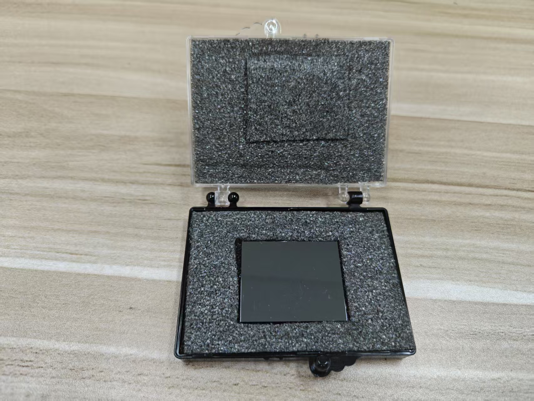
Cadmium Zinc Telluride (CZT) detectors are widely used in SPECT-CT for their high stopping power, direct conversion mechanism, and excellent energy and spatial resolution. However, the material quality of CZT crystals significantly affects detector performance. One of the primary defects in CZT crystals is tellurium (Te) inclusions—microscopic clusters of excess Te formed during crystal growth. These inclusions act as localized regions of distorted lattice structure, introducing charge trapping and scattering centers that influence both the energy resolution and image quality. Understanding the role of Te inclusion density is critical for optimizing CZT detectors in clinical imaging.
## Formation and Characteristics of Te Inclusions
Te inclusions arise during the Bridgman or vertical gradient freeze growth of CZT crystals due to slight deviations in stoichiometry or non-uniform cooling rates. These inclusions typically range from sub-micron to tens of microns in size and are dispersed throughout the crystal. Higher inclusion density correlates with a greater number of localized defects. Since the charge transport properties in CZT are sensitive to lattice uniformity, these inclusions create non-uniform electric fields, increase the probability of electron and hole trapping, and induce fluctuations in charge collection efficiency. The size, shape, and distribution of Te inclusions therefore directly affect detector performance metrics such as energy resolution and signal-to-noise ratio.
## Impact on Charge Transport and Energy Resolution
Energy resolution in CZT detectors depends on the accurate measurement of the total charge generated by a gamma photon interaction. Te inclusions act as trapping centers for both electrons and holes, reducing the collected charge and introducing variability in pulse amplitude for identical energy photons. The cumulative effect is broadening of the photopeak, manifesting as degraded energy resolution. In clinical SPECT-CT, this reduction in energy resolution diminishes the detector's ability to discriminate between primary photons and scattered events or to separate photopeaks from different isotopes. Consequently, higher Te inclusion density leads to increased spectral overlap, reduced scatter rejection, and potential contamination between isotopes during multi-isotope imaging.
## Spatial Variability and Image Uniformity
In addition to spectral effects, Te inclusions introduce spatial non-uniformities in detector response. Pixels aligned with regions of higher inclusion density may exhibit reduced charge collection efficiency, while pixels in lower-density areas perform optimally. This variation can lead to spatial artifacts in reconstructed images, including streaking, local count deficits, or apparent inhomogeneities in radiotracer distribution. Such non-uniformities can compromise quantitative measurements in SPECT-CT, where accurate voxel-level activity mapping is essential for assessing perfusion, receptor density, or tumor uptake.
## Effect on Count Statistics and Sensitivity
The presence of Te inclusions can reduce effective detector sensitivity by causing partial charge loss or complete rejection of events that fail energy threshold criteria. In practical terms, more photons are excluded from the final dataset due to misclassified energies, resulting in lower count statistics. Reduced photon counts increase statistical noise, degrading image quality, and requiring longer acquisition times to achieve clinically acceptable image quality. High inclusion density thus imposes a trade-off between crystal size and usable detector area versus overall image fidelity and acquisition efficiency.
## Mitigation Strategies in Detector Design
Several strategies are employed to mitigate the effects of Te inclusions in CZT SPECT-CT detectors. First, careful material selection and crystal growth optimization reduce inclusion density. Techniques such as slow cooling, stoichiometric control, and post-growth annealing minimize the formation of Te clusters. Second, detector design incorporates small pixel pitches and segmentation, allowing for localized charge collection and the use of pixel-specific calibration factors to correct for inclusion-related variations. Advanced signal processing algorithms can also identify partial charge events associated with inclusions and apply energy or position corrections, partially restoring energy resolution and uniformity.
## Calibration and Quality Control
Effective calibration is essential to compensate for residual Te inclusion effects. Flood-field calibration maps per-pixel sensitivity variations, enabling correction of both energy and spatial non-uniformities. Energy calibration routines ensure that pixels affected by inclusions still align their photopeak positions with reference energies. Additionally, quality control procedures, including imaging uniform phantoms and monitoring spectral broadening, allow manufacturers and clinical teams to quantify inclusion-related performance impacts and validate detector suitability for routine SPECT-CT imaging.
## Clinical Implications
High Te inclusion density can directly affect the diagnostic quality of SPECT-CT scans. Poor energy resolution may reduce the ability to reject scattered photons, impair multi-isotope discrimination, and introduce artifacts that mimic or obscure pathological uptake. Spatial non-uniformities can distort functional maps, particularly in small or low-contrast lesions. Conversely, low inclusion density CZT crystals provide more consistent charge collection, sharper photopeaks, and improved uniformity, enabling accurate quantitative imaging, better lesion detectability, and higher confidence in clinical interpretations.
## Conclusion
Te inclusion density in CZT crystals is a critical determinant of detector performance in SPECT-CT. High inclusion density degrades energy resolution, introduces spatial non-uniformities, reduces effective sensitivity, and can compromise image quality and quantitative accuracy. Mitigation strategies, including optimized crystal growth, pixelated detector design, advanced signal processing, and rigorous calibration, are essential to minimize the impact of inclusions. Maintaining low Te inclusion density ensures that CZT detectors achieve their full potential for high-resolution, quantitative SPECT-CT imaging, supporting reliable diagnostic and functional assessments across cardiac, neurological, and oncological applications.
CdZnTe Association (CdZnTe.com)
https://www.cdznte.com/blog/how-does-te-inclusion-density-in-czt-material-influence-energy-resolution-and-image-quality-in-spect-ct-using-a-czt-detector.html