## Introduction to Stopping Power in CZT Detectors
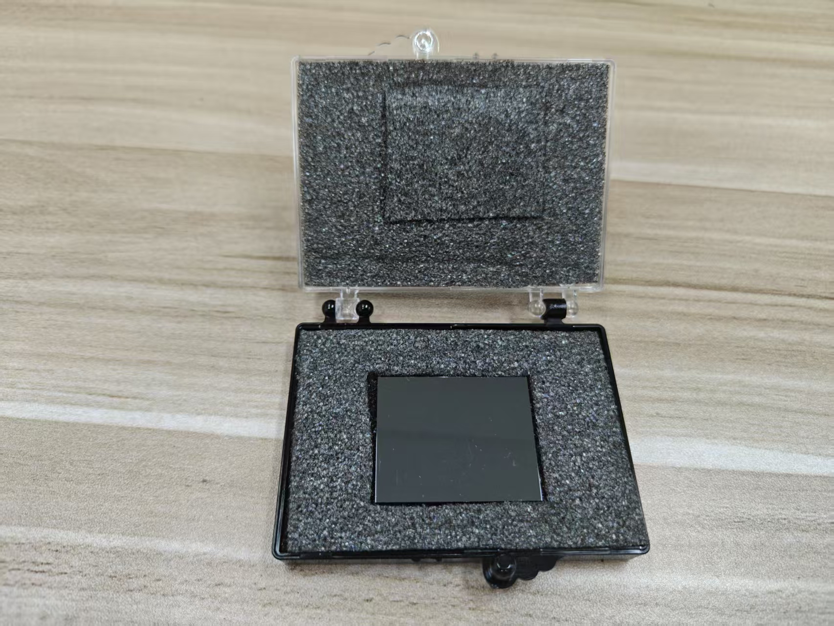
Cadmium Zinc Telluride (CZT) detectors are widely utilized in modern medical imaging systems such as SPECT-CT due to their unique combination of high atomic numbers (Cd: 48, Zn: 30, Te: 52), high density (~5.8 g/cm³), and direct conversion capability. Stopping power refers to a material’s ability to absorb incoming gamma photons and convert them into measurable electrical signals. In medical imaging, higher stopping power translates directly into higher photon detection efficiency, which is critical for achieving high sensitivity, especially under low-dose imaging protocols or high-speed acquisitions. The superior stopping power of CZT provides a distinct advantage over conventional NaI(Tl) scintillators in terms of detection efficiency, signal fidelity, and quantitative imaging performance.
## Mechanisms Contributing to High Stopping Power
The high stopping power of CZT arises from its material properties:
*
Photoelectric effect dominance: The high atomic numbers of Cd, Zn, and Te enhance photoelectric absorption, particularly for low- to medium-energy gamma photons (70–150 keV), which are commonly used in SPECT imaging.
*
Increased density: Greater electron density increases the probability of photon interactions per unit volume, improving overall detection efficiency.
*
Thicker detector volumes achievable without significant signal loss: The combination of high density and direct conversion allows fabrication of detectors with sufficient thickness to absorb a large fraction of incident photons without compromising spatial resolution.
*
Reduced Compton escape: High-Z materials reduce the probability that a photon will scatter out of the detector volume before depositing its full energy, enhancing photopeak detection.
These mechanisms collectively ensure that a higher proportion of emitted photons from the patient are detected, improving the system’s intrinsic sensitivity.
## Impact on Sensitivity in Medical Imaging
Higher stopping power directly affects sensitivity—the ability to detect a given number of photons per unit administered activity:
*
Increased photon capture: More photons are absorbed and converted into measurable charge, reducing the number of missed events and improving counting statistics.
*
Improved low-dose imaging: Enhanced sensitivity allows imaging protocols to reduce radiopharmaceutical activity while maintaining sufficient counts for diagnostic quality.
*
Shorter acquisition times: With higher photon collection efficiency, fewer rotations or shorter scan durations are needed, improving patient comfort and throughput.
*
Enhanced small lesion detectability: Higher sensitivity reduces statistical noise in reconstructed images, allowing subtle lesions to be distinguished from background activity.
Overall, the improved stopping power allows the imaging system to capture a more complete representation of the photon flux, which is particularly important in pediatric imaging, oncology, and cardiac studies where low-dose or rapid acquisition is required.
## Synergy with Direct Conversion Mechanism
CZT’s direct conversion of gamma photons to charge carriers maximizes the benefit of high stopping power:
*
Immediate charge collection: Every absorbed photon contributes directly to the signal without intermediate light conversion, reducing loss mechanisms inherent in scintillator-PMT systems.
*
High-energy fidelity: Complete absorption of photon energy enhances spectral resolution, allowing more precise energy discrimination and reducing scatter contamination.
*
Integration with pixelated electrodes: The combination of high stopping power and fine pixelation maintains spatial resolution even at thicker detector volumes, avoiding blurring while capturing more photons.
The synergy of material stopping power and direct conversion ensures that high intrinsic sensitivity is fully realized in clinical imaging scenarios.
## Influence on Quantitative Accuracy
Higher photon capture improves quantitative metrics:
*
Better statistical reliability: More detected counts per voxel reduce Poisson noise, allowing more precise activity measurements.
*
Enhanced energy-based corrections: High photopeak efficiency improves the accuracy of scatter and attenuation corrections, as more photons are detected in their correct energy bins.
*
Support for multi-isotope imaging: With higher sensitivity, the system can resolve multiple isotopes simultaneously without compromising count statistics for each radionuclide.
These effects are critical for accurate assessment of tracer uptake in perfusion studies, tumor imaging, or therapy response monitoring.
## Clinical Advantages of High Stopping Power
The practical benefits in patient care include:
*
Dose reduction potential: Lower administered radiotracer activity is feasible without sacrificing image quality.
*
Faster scans: High sensitivity allows shorter acquisition times, reducing motion artifacts and patient discomfort.
*
High-resolution imaging of small structures: Thicker CZT detectors absorb more photons while preserving intrinsic spatial resolution, enabling detailed visualization of fine anatomical structures.
*
Improved diagnostic confidence: Enhanced count statistics and reduced image noise facilitate accurate detection and characterization of lesions, particularly in low-contrast or small-volume studies.
These clinical benefits demonstrate the direct impact of CZT stopping power on workflow efficiency, patient safety, and diagnostic accuracy.
## Considerations and Optimization
While high stopping power is advantageous, it must be balanced with system design:
*
Detector thickness vs. charge collection: Excessive thickness may increase charge trapping or depth-dependent effects, requiring depth-of-interaction corrections to maintain spatial resolution.
*
Pixelation and electronic readout: High sensitivity must be paired with fine pixel readout and low-noise electronics to fully exploit the additional counts.
*
Thermal management: Higher photon capture increases total charge collection, which can stress electronics if not adequately designed for thermal stability.
Careful system engineering ensures that the high stopping power translates into tangible sensitivity gains without introducing secondary artifacts or electronic limitations.
## Conclusion
The superior stopping power of CZT detectors is a fundamental factor in achieving higher sensitivity in medical imaging. By efficiently absorbing gamma photons through photoelectric interactions and high-density material properties, CZT ensures that a larger fraction of emitted radiation is detected and converted into measurable charge. This high intrinsic efficiency allows for lower administered doses, faster scan times, improved lesion detectability, and more accurate quantitative imaging. When combined with direct conversion, pixelated architecture, and advanced electronics, CZT detectors leverage their stopping power to deliver significant clinical advantages in SPECT-CT and other nuclear medicine applications.
CdZnTe Association (CdZnTe.com)
https://www.cdznte.com/blog/how-does-the-excellent-stopping-power-of-a-czt-detector-contribute-to-higher-sensitivity-in-medical-imaging.html